A New Antigen Test Method for COVID-19: Achieving Rapid, High-accuracy Detection with Electron Microscope
May. 13, 2022
Daishi Kawabata
Over two years have passed since the outbreak of the COVID-19 pandemic. As waves of increasing numbers of infected people strain the capacities of medical facilities, technology that can test for viruses in a short time and with high accuracy is required.
To meet such needs, in February 2022, Hitachi Social Information Services partnered with Hamamatsu University School of Medicine and other organizations to develop a new antigen test method. By combining regular commercially available antigen test kits and a desktop scanning electron microscope, the new method achieves detection accuracy as high as PCR test levels in the short time of about 30 minutes. In the future, this technology is also expected to find broad application in the testing of other infectious diseases in addition to COVID-19.
Two types of tests for COVID-19
There are two types of tests for determining infection by COVID-19: the PCR test and the antigen test. Currently, the main test in use is the PCR test, which employs special equipment to detect the genetic material of the virus in a sample taken from the nasal cavity. Boasting high detection accuracy, the test has been adopted by most medical institutions. However, as pointed out by Associate Professor Hideya Kawasaki of Hamamatsu University School of Medicine, there is an issue of testing time.
"Although the PCR test has achieved high accuracy, it goes through a process of the amplification of viral genetic material, resulting in test times of at least two to four hours. Moreover, only special facilities have the device used for detection, necessitating the time to transport samples there. Therefore, depending on the region, it could take one to several days, from sample acquisition to test results."
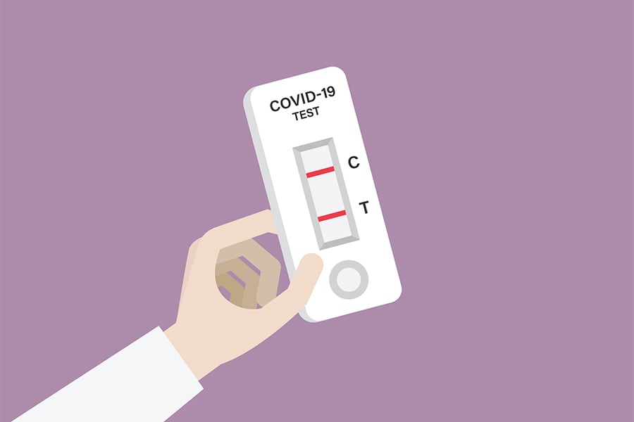
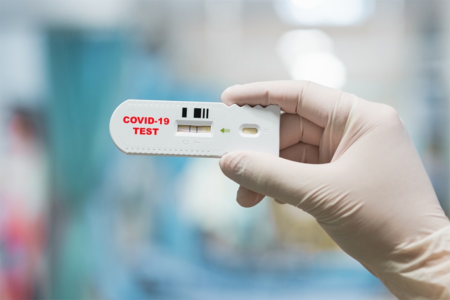
The antigen test on the other hand, looks for a type of protein, known as an antigen, that is specific to the virus. A positive or negative test result is determined by dropping mucus taken from the nasal mucosa onto a commercially available test kit and observing its test line. Antigen test usage is increasing becoming popular in Japan because it is easy to use and delivers rapid results in approximately 15 minutes.
However, the antigen test kits have the disadvantages of lower accuracy compared to the PCR test and of a high rate of false negatives (i.e., a negative result despite the presence of the virus). For this reason, the PCR test tended to be prioritized in the diagnosis of COVID-19 at medical facilities.
A new test method using "NanoSuit"
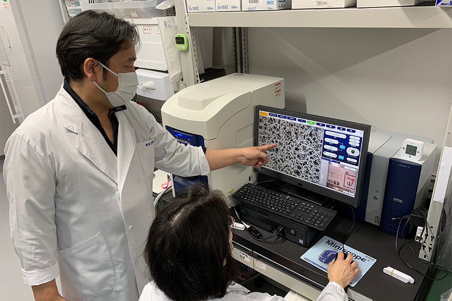
(Above left. Photo provided by Hamamatsu University School of Medicine)
Against this backdrop, Hamamatsu University School of Medicine and Hitachi Social Information Services developed a new antigen test method. Dr. Kawasaki says that the method represents an improvement on the faults of both PCR test and existing antigen test kits:
"The major advantage of the new antigen test method that we developed is that it raises detection sensitivity to PCR test levels while providing results more simply and rapidly than a PCR test."
Takumi Tandou of Hitachi Social Information Services, who was involved in development, explains that "the test method is very simple":
"First, you drop a sample, such as that from the nasal mucosa, onto a commercially available kit. After about 15 minutes, you then drop a special liquid called 'NanoSuit' onto it. Next, you just need to observe the test line under a desktop scanning electron microscope."
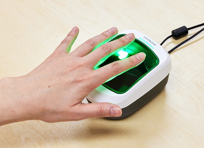
NanoSuit is a solution originally developed by Hamamatsu University School of Medicine that allows the observation of viral and other types of specimens under the vacuum conditions of an electron microscope.
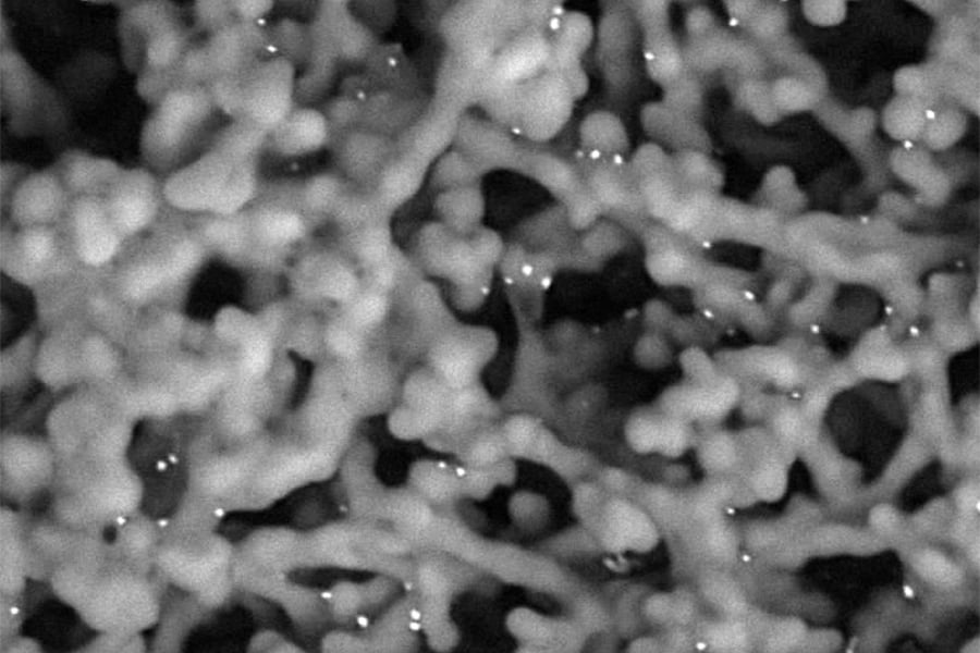
white particles (metal grains attached to the antigen) observed in the image.
If the virus is present in the dropped sample, antigen that is specific to the virus binds to antibodies attached to metal particles, which then accumulate on the test line. These metal particles appear as white dots when the test line is observed under an electron microscope. A positive result is indicated when the number of white dots exceeds a certain value.
This method raised the accuracy of virus detection to PCR test levels. Since the genetic material amplification process is not required,, it takes only about 30 minutes to get the results (i.e., the reaction time of the test kit plus the observation time under the desktop electron microscope). The new method is therefore capable of significant reductions in time compared to the PCR test.
Inspiration for a new test method

The new antigen test method achieved accuracy as high as PCR tests and has a test time of only about 30 minutes. Although expectations are high that it will find wide use in the future, the journey to this point was not a smooth one.
The beginnings of development go back to 2016. Tandou, who was then researching manufacturing equipment for semiconductors, was using an electron microscope to observe the structure of semiconductors on a daily basis. It was during this time that an opportunity arose:
"One time my kids had come down with influenza. While nursing them, I realized that since you could see semiconductors in such detail with an electron microscope, you ought to be able to see an influenza virus. The idea sprang from this."
Right away, Tandou consulted with various experts both in and outside the company regarding whether or not it was possible to observe a virus with a desktop electron microscope. However, he said that he "got mostly skeptical responses." The reason given was that a virus was too small to resolve with the magnification available on a desktop scanning electron microscope. Amid such trials & tribulations, Tandou learned of Dr. Kawasaki at Hamamatsu University School of Medicine, who was researching viruses using an electron microscope, and went to meet him.
Since then, Tandou frequently traveled from Tokyo to Hamamatsu to continue a dialogue with Dr. Kawasaki about how he "wanted to make it possible to measure the virus in the body as easily as taking body temperature with a thermometer." Looking back, Dr. Kawasaki said that it was "a single, highly impressive experience" that provided the impetus for their joint research project. Elaborating, he said:
"When I used an antigen test kit for influenza virus and, as an experiment, dropped a sample with NanoSuit, and looked through a desktop electron microscope, I was impressed at how clearly I could see the virus(precisely, the metal particles). This stimulated my interest as a doctor in the possibility of being able to create a new testing method by raising sensitivity even further."
Demonstration testing that verified high accuracy
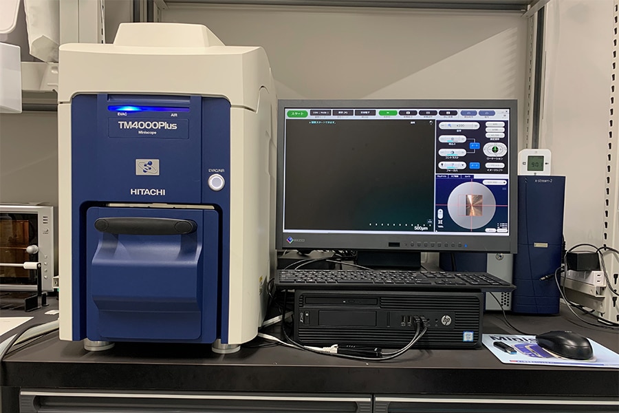
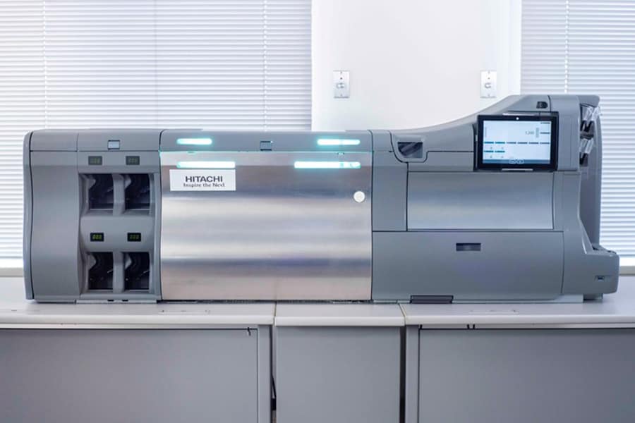
(manufactured by Hitachi High-Tech)
This eventually led to the start of a full-scale joint research by Hitachi and Hamamatsu University School of Medicine in 2019. Initially, the project was aiming to develop a test method for influenza, but then came the spread of COVID-19 pandemic. The team then shifted the target of the research to the COVID-19.
After a year of trial and error, a new antigen test method was finally completed. To verify its effectiveness, demonstration testing was carried out from December 2020 to October 2021. A total of 88 clinical samples was taken from the saliva and nasal mucous membranes of 45 people suspected of having contracted COVID-19. The team compared the results from a PCR test, a conventional antigen test, and the new desktop electron microscope-based test method.
It was found that, among samples determined to be positive by the PCR test, only 43.3% could be diagnosed as positive by the conventional antigen test kits. In contrast, 86.7% were diagnosed as positive by the desktop electron microscope-based antigen test. These results indicate a drastic improvement in sensitivity, which is said to be equivalent to a maximum of about 500 to 1,000 times that of testing that relies on conventional antigen test. This shows that the newly developed test method can be expected to significantly improve test accuracy.
Dr. Kawasaki cites the following as the most-important point:
"When we carried out the new test method on samples that were determined positive by a PCR test and negative by an antigen test kit, our results were 73.3% positive. This difference in test sensitivity is overwhelming. This value shows that the new method has testing performance equivalent to PCR diagnosis. We can say that we achieved the highest level of sensitivity for antigen testing."
Prospects for the new antigen test method
Tandou is now working on further development aimed at implementing the new testing method. He says:
"We are currently developing image analysis technology that uses AI to automatically count the number of virus particles, and are working to automate and improve the test speed."
Additionally, Dr. Kawasaki also expresses high hopes for future possibilities:
"I believe that introducing this test method as a service that can be easily used by anyone, even after the COVID-19 pandemic, will drastically reduce the load on doctors and public health nurses nationwide—and even on patients themselves. As long as they have a desktop scanning electron microscope, schools and workplaces will also be able to perform tests and use them in regular health check-ups of students and staff."
Immunochromatography—the technology used in antigen tests—is widely employed in COVID-19, influenza, pregnancy, and other types of tests. And in addition to being applied to testing for infectious disease in livestock, it is also used in testing for food allergens and agricultural chemicals residue. Tandou says that "The test method that we developed is opening the way to raise the sensitivity of a variety of tests by using an electron microscope."
Going forward, Hitachi will continue to explore further possibilities for application of this testing method in the post-COVID-19 world.