Finally – Accurate, Efficient and Comfortable Breast Ultrasound
SOFIA 3D breast ultrasound system solves issues for women with dense breast tissue
On October 1, 2009, Governor Jody Rell of Connecticut, a breast cancer survivor, signed a bill requiring communications be sent to patients when breast density was noted during their mammography examination. The goal of the bill was to aid in early detection of breast cancer. Following that Connecticut bill, 31 states now have some type of breast density notification legislation. The legislation raises awareness about how dense breasts, which have high proportions of glandular and fibrous tissue, can make it harder for radiologists to see cancer.
Breast density is one of the strongest predictors of the failure of mammography screening alone to detect cancer. A full 40% of the women who have mammograms have dense breasts. However, many women don't know their breast density and what that may mean when it comes to their risk of getting breast cancer.

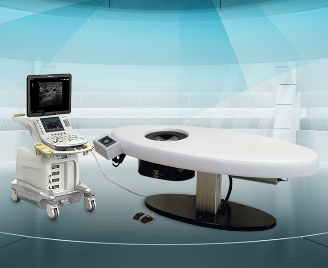
SOFIA 3D breast ultrasound system
Studies have shown that mammography alone is significantly less accurate in this subset of women. A study facilitated by the American College of Radiology (ACR) showed that adding a supplemental ultrasound exam in these patients can increase the number of cancers detected by 55%.1 While the adoption of whole-breast ultrasound was initially slow, state laws requiring notification and many recent articles on the subject have increased awareness of the issue leading more women to talk to their doctors about adjunctive imaging options like ultrasound. In New Jersey, the number of supplemental breast ultrasounds performed, increased by 651% in the 18 months following the passage of their law as compared to the 18-months prior.2
The American Roentgen Ray Society (ARRS), the first and oldest radiology society in the United States, is also aware of this issue. At their meeting in May 2017, researchers from Elizabeth Wende Breast Care in Rochester, NY, presented their findings that ultrasound outperformed digital breast tomosynthesis (DBT) in women with heterogeneously or extremely dense tissue.3 For women with dense breast tissue, this information can lessen anxiety and confusion and increase confidence in their choices.
Designed for comfort, designed for women
While mammography has been the most accurate way to predict breast cancer, it can produce anxiety in women with dense breast tissue which is compounded by the discomfort many women experience with an exam. Some whole-breast ultrasound system designs cause similar discomfort by forcing women to lie on their backs with breasts exposed during the exam while a substantial amount of ultrasound gel is continually reapplied. Other designs have scanning plates that use external mechanical pressure to compress the breast, in effect pinning patients to the table. None of these systems have been designed with the comfort and privacy that women need.
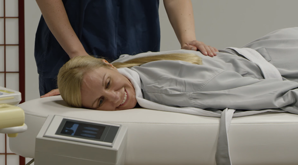
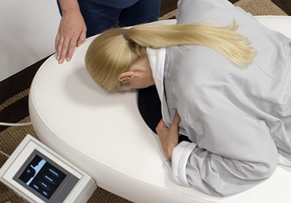
In contrast, Hitachi's SOFIA™ 3D breast ultrasound system is designed to offer a private and comfortable experience. SOFIA's multi-layer memory foam surface provides a comfortable environment during the quick exam. The patient is scanned in the prone position and remains covered during the exam, increasing privacy. The patient's own body weight naturally provides compression, so the patient can gauge her own comfort. Ergonomic design considerations for patients are top of mind for Hitachi.
Hitachi’s SOFIA offers many efficiencies to ease its adoption into a providers existing practice. For example, SOFIA is able to deliver scan times of 30 seconds per breast. In fact, bilateral whole-breast exams can be scheduled in 10 minute intervals. In comparison, handheld bilateral breast exams may take up to 30-45 minutes per exam. Ease of use for operators is another efficiency that the SOFIA system offers. SOFIA's user interface touch screen provides operators with an intuitive icon driven menu. One button launches a fully automated scan procedure providing rapid whole-breast image acquisition.
SOFIA also provides flexible options to conserve exam room resources. SOFIA does not require a dedicated room for whole-breast ultrasound. When the space is not being used for whole-breast imaging, SOFIA’s scan table can be used as a conventional scan bed for a wide variety of diagnostic ultrasound exams. Unlike other automated breast ultrasound systems, SOFIA requires no additional consumables and no time-consuming pre-scan preparation.
In terms of reimbursement, CMS released a new CPT code in 2015 which directly impacted bilateral breast exams. As a result, reimbursement for this exam increased almost 64%. SOFIA providers have not run into any significant situations where private payers are not reimbursing for this code—even in states without notification or insurance laws.
Driving Social Innovation in U.S. healthcare
When combined with mammography, The SOFIA 3D breast ultrasound system gives women with dense breast tissue a new way to detect breast cancer. It also solves many of the economic and logistical challenges associated with whole-breast ultrasound by using a full-field radial scanning method that delivers fast scanning and image interpretation times. The throughput efficiencies and advanced patient comfort make SOFIA the ideal solution for radiology practices that are expanding solutions for women with dense breasts.
Co-creating solutions to address health challenges is at the heart of social innovation. Cancer has become a leading cause of death, especially in developed countries. Advancing the science of cancer care and supporting education reflects Hitachi’s goal of social innovation. Hitachi Social Innovation Business seeks to play a vital role in the diagnosis and treatment of disease while co-creating solutions that improve healthcare in the U.S. Hitachi recognizes that healthcare is an essential part of social infrastructure to support human life in the 21st century.


